WE ACCEPT MOST INSURANCE PROVIDERS!
Few diagnostic tools in the world of cardiovascular medicine hold as much significance and versatility as the echocardiogram. This innovative technology has revolutionized the way we assess heart health, offering unparalleled insights into cardiac structure, function, and overall performance.
For those unfamiliar with this groundbreaking innovation, we’ll provide a general overview on this page. If you have any particular questions or concerns, you are welcomed to call Capitol Cardiology Associates to schedule an appointment with a licensed cardiologist. We are also happy to arrange a walk-in for as soon as possible.
An echocardiogram, often referred to as an echo, is a non-invasive imaging test that utilizes ultrasound waves to create real-time images of the heart. Unlike traditional X-rays or CT scans, echocardiograms provide dynamic views of the heart's chambers, valves, blood flow patterns, and overall cardiac function.
This comprehensive assessment allows doctors to evaluate heart health, detect abnormalities, monitor disease progression, and guide treatment strategies with remarkable precision. It is a must-have tool in any holistic approach to cardiovascular medicine.
Echocardiograms offer a comprehensive assessment of cardiac anatomy and function. It allows us to learn measurements of chamber sizes, valve function, wall motion abnormalities, and ejection fraction (a key indicator of heart pumping efficiency).
Unlike invasive procedures such as cardiac catheterization, echocardiograms are non-invasive and pose minimal risk to patients. They do not involve radiation exposure, making them safe for repeated use, even in sensitive populations. Furthermore, echocardiography provides real-time imaging, which allows healthcare providers to visualize heart structures and blood flow dynamics as they occur. This real-time feedback is essential for immediate clinical decision-making. By providing detailed insights into cardiac function and pathology, echocardiograms guide treatment decisions, surgical interventions, and ongoing management strategies for a wide range of cardiovascular conditions.
Before the echocardiogram, there is typically minimal preparation required. Patients may be asked to avoid eating or drinking for a few hours before the test, particularly for transesophageal echocardiography (TEE) procedures that involve inserting a probe into the esophagus.
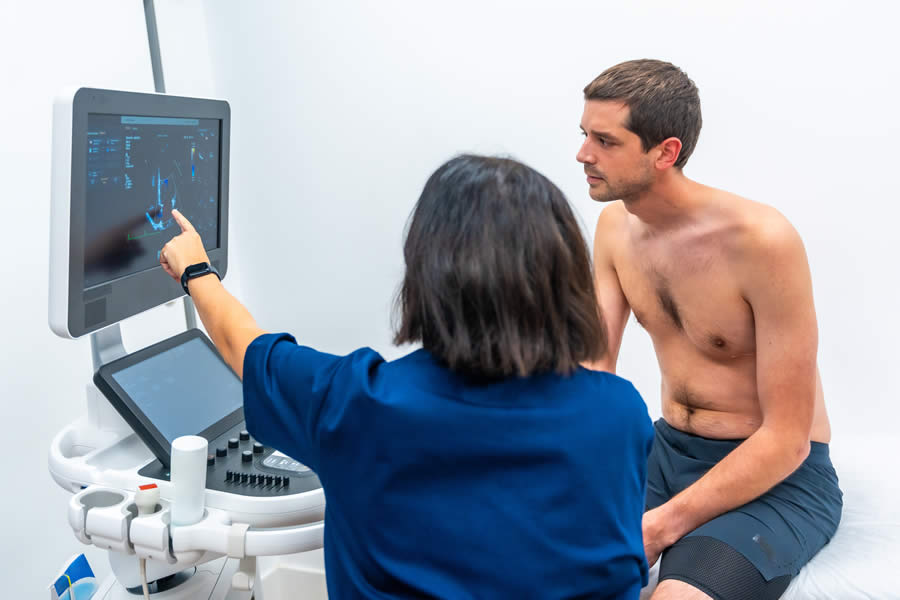
During a transthoracic echocardiography (TTE), the patient lies comfortably on an examination table, while a transducer (probe) is placed on the chest to capture images. For a TEE, a specialized probe is inserted through the mouth or nose into the esophagus for clearer imaging of certain heart structures.
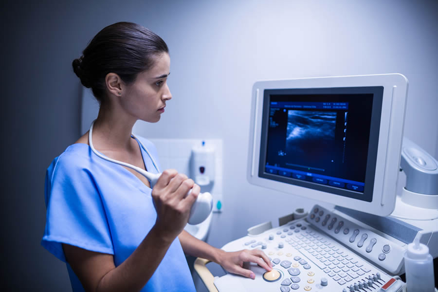
Next up is the ultrasound imaging. The echocardiographer moves the transducer across specific areas of the chest (or esophagus) to obtain different views of the heart. The ultrasound waves create detailed images displayed on a monitor in real time.
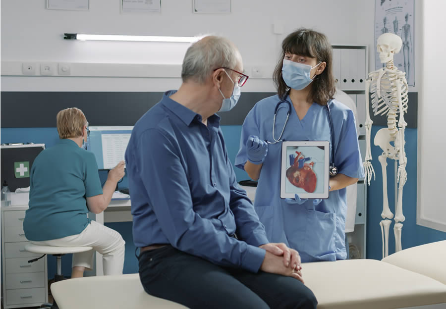
The echocardiographer interprets the images, assessing cardiac structures, valve function, blood flow patterns, and any abnormalities. Once the echocardiogram is complete, patients can typically resume their normal activities without any restrictions. Results are reviewed by an experienced cardiologist, who integrates them into the patient's overall clinical assessment and treatment plan.
If you have any scares or concerns regarding your heart health, it is always advisable to see a licensed cardiologist sooner rather than later. Identifying issues and vulnerabilities can be literal lifesavers. Residents in the area can call Capitol Cardiology Associates to schedule an appointment with a heart doctor. Our team is on standby to take your call today.